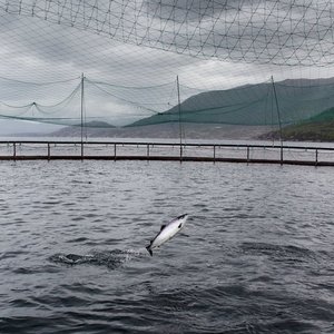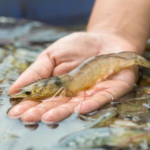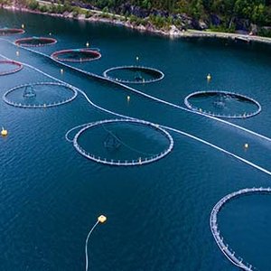
The Danish Technological Institute (DTI), in collaboration with BioMar, investigated batches of fish larvae after being fed different experimental diets. BioMar was interested in how the experimental diets affected the digestion process of fish, and the company found great potential in non-destructive 3D imaging as a complement to dissection and histological analysis.
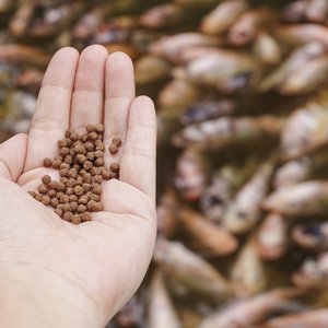
“Our collaboration with DTI has tremendously aided in expanding our research tools to increase our knowledge of fish physiology. The overarching gain to include this top-of-the-art technology is to continue improving what we offer to the aquaculture industry in the form of a feed. In other words, each pellet we produced is based on solid science, and this exciting collaboration provided us with the right tools,” said Pedro Gómez, senior scientist, Biomar Denmark.
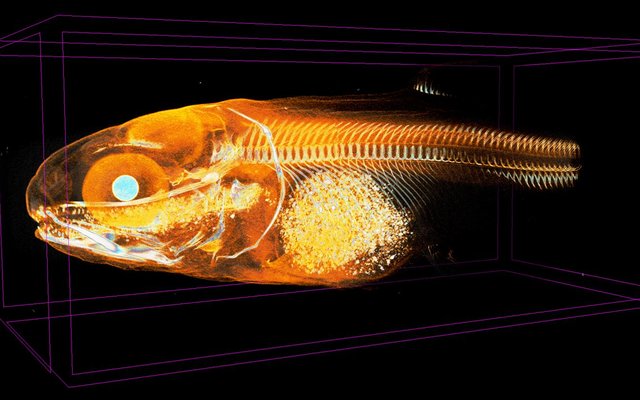
3D scans were first performed on a laboratory Micro-CT available at DTI, a procedure which, for this type of specimen, includes heavy metal staining to increase X-ray absorption in the low-density tissue. A collaboration with the European Synchrotron Radiation Facility (ESRF) in France was then initiated, which led to the project continuing with their new state-of-the-art imaging technique, HiP-CT (Hierarchical Phase-Contrast Tomography). Data quality was drastically improved with the introduction of phase contrast-CT, and heavy metal staining was no longer necessary. The much-improved level of detail and contrast seen in the soft tissues of the fish was made possible by the phase contrast and high flux achieved at the synchrotron facility.
“This is the most jaw-dropping data I've ever worked on! Many of our customers come from the food or pharmaceutical industry, and I can easily see how they could benefit from this type of analysis,” said Erik Wisaeus, business manager at DTI.
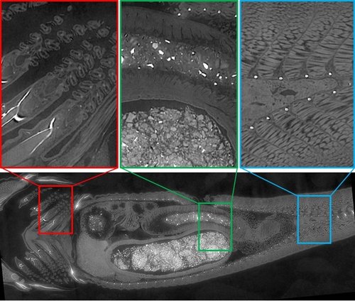
The collage below shows detailed images from a single virtual cross-section of an unstained small fish of ca 25 mm in length. The insets show an incredible level of detail, here exemplified in a region of the gills, the stomach/intestine, and an area near the rectum. The collected data makes it possible to study how different experimental diets are processed by fish. Apart from a series of cross-section images, a variety of 3D visualizations were produced to be able to compare the various specimens.
Phase contrast-CT is a new tool to see unprecedented detail in soft tissues and other organic materials and will revolutionize how we analyze food products, medical products and tissue for research and product development, researchers said. The highest resolution that can be reached using phase contrast-CT at a synchrotron facility is 100 times that of a medical CT scanner, and contrary to regular CT scanning, good contrast can now be achieved also in soft tissue and other low-density materials.